Heart

The heart is a myogenic muscular organ found in all animals with a circulatory system (including all vertebrates), that is responsible for pumping blood throughout the blood vessels by repeated, rhythmic contractions. The term cardiac (as in cardiology) means "related to the heart" and comes from the Greek καρδιά, kardia, for "heart".

The vertebrate heart is composed of cardiac muscle, which is an involuntary striated muscle tissue found only in this organ, and connective tissue. The average human heart, beating at 72 beats per minute, will beat approximately 2.5 billion times during an average 66 year lifespan, and weighs approximately 250 to 300 grams (9 to 11 oz) in females and 300 to 350 grams (11 to 12 oz) in males.
In invertebrates that possess a circulatory system, the heart is typically a tube or small sac and pumps fluid that contains water and nutrients such as proteins, fats, and sugars. In insects, the "heart" is often called the dorsal tube and insect "blood" is almost always not oxygenated since they usually respirate (breathe) directly from their body surfaces (internal and external) to air. However, the hearts of some other arthropods (including spiders and crustaceans such as crabs and shrimp) and some other animals pump hemolymph, which contains the copper-based protein hemocyanin as an oxygen transporter similar to the iron-based hemoglobin in red blood cells found in vertebrates.
The human heart has a mass of between 250 and 350 grams and is about the size of a fist. It is located anterior to the vertebral column and posterior to the sternum.
It is enclosed in a double-walled sac called the pericardium. The superficial part of this sac is called the fibrous pericardium. This sac protects the heart, anchors its surrounding structures, and prevents overfilling of the heart with blood.

The outer wall of the human heart is composed of three layers. The outer layer is called the epicardium, or visceral pericardium since it is also the inner wall of the pericardium. The middle layer is called the myocardium and is composed of muscle which contracts. The inner layer is called the endocardium and is in contact with the blood that the heart pumps. Also, it merges with the inner lining (endothelium) of blood vessels and covers heart valves.
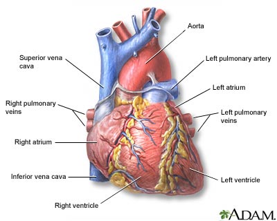
The human heart has four chambers, two superior atria and two inferior ventricles. The atria are the receiving chambers and the ventricles are the discharging chambers. The right ventricle discharges into the lungs to oxygenate the blood. The left ventricle discharges its blood toward the rest of the body via the aorta.
The pathway of blood through the human heart consists of a pulmonary circuit and a systemic circuit. Blood flows through the heart in one direction, from the atria to the ventricles, and out of the great arteries, or the aorta for example. This is done by four valves which are the tricuspid valve, the mitral valve, the aortic valve, and the pulmonary valve.
In amphibians and most reptiles, a double circulatory system is used but the heart is not completely separated into two pumps. The development of the double system is necessitated by the presence of lungs which deliver oxygenated blood directly to the heart.
In living amphibians, the atrium is divided into two separate chambers by the presence of a muscular septum even though there is only a single ventricle. The sinus venosus, which remains large in amphibians but connects only to the right atrium, receives blood from the vena cavae, with the pulmonary vein by-passing it entirely to enter the left atrium.
In the heart of lungfish, the septum extends part-way into the ventricle. This allows for some degree of separation between the de-oxygenated bloodstream destined for the lungs and the oxygenated stream that is delivered to the rest of the body. The absence of such a division in living amphibian species may be at least partly due to the amount of respiration that occurs through the skin in such species; thus, the blood returned to the heart through the vena cavae is, in fact, already partially oxygenated. As a result, there may be less need for a finer division between the two bloodstreams than in lungfish or other tetrapods. Nonetheless, in at least some species of amphibian, the spongy nature of the ventricle seems to maintain more of a separation between the bloodstreams than appears the case at first glance. Furthermore, the conus arteriosus has lost its original valves and contains a spiral valve, instead, that divides it into two parallel parts, thus helping to keep the two bloodstreams separate.
The heart of most reptiles (except for crocodilians; see below) has a similar structure to that of lungfish but, here, the septum is generally much larger. This divides the ventricle into two halves but, because the septum does not reach the whole length of the heart, there is a considerable gap near the openings to the pulmonary artery and the aorta. In practice, however, in the majority of reptilian species, there appears to be little, if any, mixing between the bloodstreams, so the aorta receives, essentially, only oxygenated blood.


0 comments: